 Collaborators: Naveen Nagarajan, PhD, Mario Capecchi, PhD
Department: Human Genetics
Project
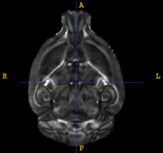
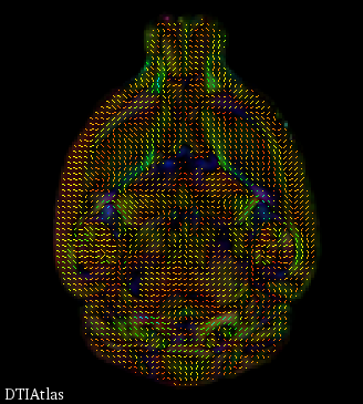
Obsessive Compulsive disorder (OCD) is a devastating anxiety disorder that is characterized by recurrent, unwanted thoughts (obsessions) and/or repetitive behaviors (compulsions). To understand the neurobiological underpinnings of OCD and to develop innovative therapeutic approaches, we have developed a mouse model that exhibits some of the hallmarks of this disorder. Deletion of Hoxb8, a homeobox domain gene leads to excessive grooming behavior, hairless patches, irreversible and self-inflicted open skin lesions that are strictly similar to compulsive hair removal observed in humans. Very little is currently known about the specific brain regions and neural circuits that underlie grooming behavior. In order to better understand the functional connectivity differences between control and HoxB8 knockout mice, fMRI analysis, Magnetic resonance imaging and Diffusion tensor imaging analysis are being carried out at the Imaging facility. While Magnetic Resonance Imaging is a technique aimed to provide detailed information of normal and diseased anatomy for medical research and has become a significant imaging modality in clinical diagnosis and brain studies, Diffusion Tensor imaging provides a unique source of information about the underlying tissue structure of brain white matter including both the geometry of major fiber bundles as well as quantitative information about tissue properties represented by derived tensor measures. This project will aim to analyze group differences between wild-type (WT) and knock-out (KO) brains via structural MRI and DTI imaging.
BIDAC Contact: Clement Vachet
Progress
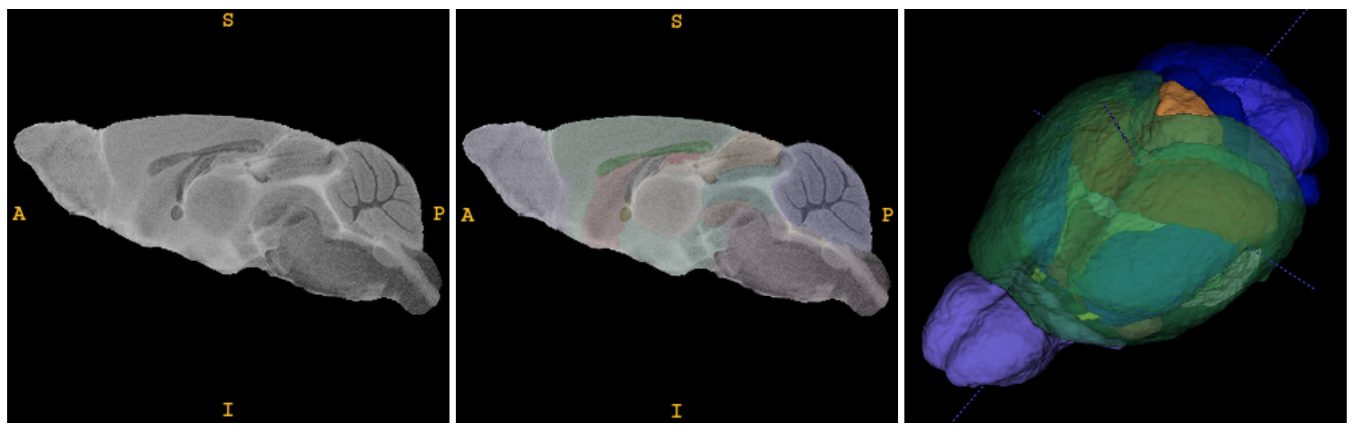
In collaboration with the Small Animal Imaging Core, we helped optimized MRI and DTI acquisitions for image analysis needs. We adapted image processing frameworks from human imaging to small animal imaging, and analyzed brain differences between WT and KO groups (9 specimen each) focusing on volumetric measurements via structural MRI and on ROI-based diffusion properties via DTI imaging. A scientific paper is currently in review in Journal Neuron.
General contribution
We developed joint expertise and Utah HSC capabilities for mouse image acquisition and analysis.
Presentations
Mouse Study: DTI analysis (.pdf)
Mouse Study: Structural MRI analysis (.pdf)
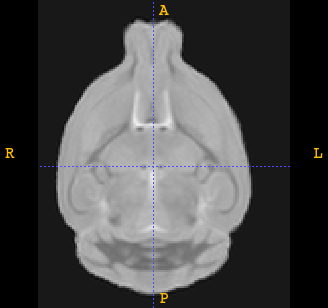
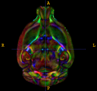
| B0 | colorFA | FA | tensors | 3D parcellation |
 |
 |
 |
 |
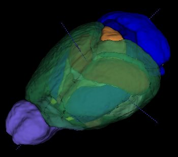 |
DTI atlas |